Keratoconus Treatment and Management Options in Auckland
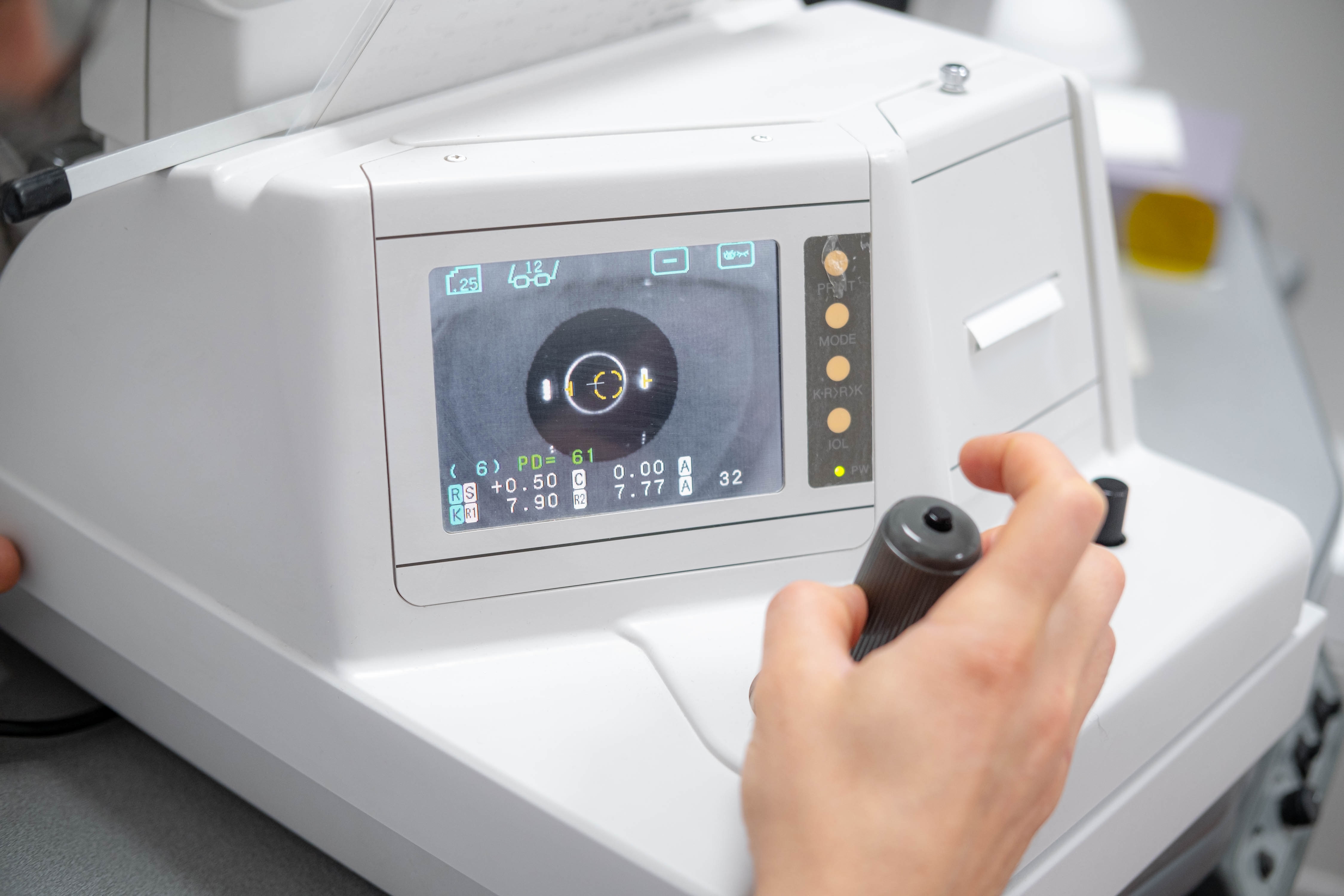
Keratoconus management in Auckland involves two parallel aims — improving vision through glasses or specialty contact lenses, and monitoring or controlling corneal progression through clinical assessment and, where appropriate, ophthalmology referral. No single pathway suits every patient; management depends on corneal shape, prescription, progression, and individual tolerance.
Keratoconus affects each patient differently. For some, the changes are gradual and vision remains manageable with glasses for years. For others, the cornea becomes irregular enough that standard correction no longer provides adequate clarity. The condition can also progress at different rates depending on age, the severity of corneal thinning, and other individual factors — which is why management cannot follow a fixed sequence.
Our keratoconus treatment in Auckland includes detailed assessment, corneal mapping, ongoing monitoring, and individually fitted specialty contact lenses. We provide optometric keratoconus care and specialty contact-lens fitting at our Rosedale practice. We do not perform corneal cross-linking, corneal transplantation, or other eye surgery — but if our findings indicate that a surgical evaluation is needed, we can arrange a referral to an ophthalmologist.
Why Keratoconus Management Is Individual
The most appropriate management depends on several clinical factors: corneal shape and curvature, the degree of visual distortion, current prescription, evidence of progression, any corneal scarring, and how well a patient tolerates contact lenses. Because these variables differ considerably between patients — and can change over time — treatment decisions must be based on each person's individual measurements and clinical picture.
Keratoconus management has two central aims. The first is improving vision through glasses or contact lenses. The second is monitoring the cornea over time and, if progression is detected, considering whether further evaluation or a referral to an ophthalmologist is warranted. These aims are separate: correcting vision with glasses or contact lenses does not prevent the cornea from continuing to change shape, and monitoring alone does not correct existing visual distortion.
How Is Keratoconus Assessed at NVISION Eyecare?
A keratoconus assessment at our practice examines several aspects of vision and corneal health. This typically includes prescription and visual acuity testing, direct examination of the cornea, and a review of previous prescriptions or measurements where available. The information gathered helps our optometrists evaluate how keratoconus is currently affecting vision and determine which management options are most suitable.
Assessing how corneal shape has changed over time is just as valuable as understanding its current state. Comparing earlier data with new measurements helps identify patterns that a single appointment cannot reveal.
What Does Corneal Topography Show?
Corneal topography produces a detailed map of the front surface of the cornea, showing variations in shape and curvature that may not be apparent during a standard vision test. At NVISION, we use the Oculus Keratograph 5M for this purpose. Our diagnostic technology provides precise measurements that help us assess corneal irregularity and design specialty lenses based on the actual shape of each patient's eye.
The Oculus Keratograph 5M is also used during specialty contact-lens fitting to support accurate lens selection and evaluation. This level of detail matters considerably when fitting lenses to an irregular cornea, where small differences in curvature can affect both comfort and visual outcome.
How Are Measurements Used to Monitor Corneal Changes?
A single topography scan shows the corneal shape at that appointment. Comparing scans taken at different points in time is what allows our optometrists to assess whether the cornea is changing — and at what rate. This is central to keratoconus monitoring because stable day-to-day vision does not reliably indicate that the cornea is unchanged.
The monitoring schedule is not universal. It is guided by the patient's age, current measurements, reported symptoms, and how quickly the cornea appears to have changed in previous assessments. Younger patients and those with signs of active progression generally require more frequent review.
What Vision Correction Options Are Available for Keratoconus?
The vision correction options for keratoconus range from standard glasses through to custom-designed specialty lenses. The most suitable correction may change as the cornea becomes more irregular, and not every patient requires specialty lenses from the outset. Our optometrists assess each patient's current vision and corneal measurements before recommending a lens type.
Do Glasses Work for Keratoconus?
Glasses can provide adequate vision correction during the earlier stages of keratoconus, particularly when corneal irregularity is mild. As the cornea becomes more irregular, a glasses prescription may no longer correct the distortion clearly — not because the prescription is wrong, but because the irregular surface scatters light in ways that a lens in a frame cannot fully compensate for.
Standard soft contact lenses face a similar limitation. They tend to follow the irregular shape of the cornea rather than creating a smooth optical surface, which reduces their effectiveness as keratoconus advances.
How Do Rigid Gas-Permeable Lenses Help with Keratoconus?
Rigid gas-permeable (RGP) lenses maintain their shape over the cornea rather than conforming to its irregular surface. This creates a more regular optical surface that can reduce visual distortion and improve clarity compared with glasses or soft lenses. RGP lenses are oxygen-permeable, which supports corneal health during wear.
Suitability for RGP lenses depends on a number of factors: how well the lens sits on the cornea, the stability and centration of the fit, patient comfort, and the visual result achieved. Not all corneal shapes accommodate an RGP lens comfortably, particularly as keratoconus advances.
What Are Scleral Lenses and How Do They Differ for Keratoconus?
Scleral lenses are significantly larger than standard corneal lenses. Rather than resting on the cornea itself, they vault over it and rest on the sclera — the white part of the eye. The space between the lens and the cornea is filled with sterile fluid before insertion, which creates a smooth, consistent surface through which light passes. This design can improve visual clarity for patients whose corneal irregularity makes smaller lenses less effective or comfortable.
Read more about scleral lenses for keratoconus and how their fit differs from smaller corneal lenses. Lens selection is always based on individual measurements and clinical findings — scleral lenses are not automatically the best option for every patient, and they do not slow or stop keratoconus progression.
What Does a Specialty Contact Lens Fitting Involve?
Specialty contact-lens fitting is considerably more detailed than a standard contact-lens appointment. The process begins with a thorough assessment of vision, prescription, corneal health, and previous experience with glasses or lenses. Corneal topography and other measurements inform the selection or design of an appropriate trial lens.
Once a trial lens is fitted, our optometrists evaluate its position, movement, clearance over the cornea, stability, and comfort — then check the visual result with the lens in place. Adjustments to the lens design may be needed based on these findings. Patients also receive instruction in inserting, removing, cleaning, and storing their lenses safely before they leave the practice.
Follow-up assessments are a standard part of the process. More than one trial lens or adjustment may be required before arriving at a final design, and the number of appointments needed varies between patients. We do not present the first trial lens as the definitive outcome.
Why Is Monitoring Keratoconus Progression Important?
Stable vision from day to day does not confirm that the cornea is unchanged. Keratoconus can continue to progress without obvious short-term changes in how clearly a patient sees. Comparing prescriptions, corneal maps, and visual results across multiple appointments provides the clearest picture of whether the condition is advancing.
Our role at NVISION is to assess and monitor keratoconus systematically. If our measurements suggest that the cornea is progressing, we discuss these findings with the patient and consider whether an ophthalmology referral is appropriate. Monitoring itself does not halt progression, but identifying changes early creates more options for timely intervention.
What Is Corneal Cross-Linking and Who Performs It?
Corneal cross-linking is a procedure designed to strengthen corneal tissue and slow or stop the progression of keratoconus. It achieves this by creating new bonds between collagen fibres within the cornea, increasing its structural rigidity. The procedure is performed by an ophthalmologist — not by an optometrist — and is generally considered when clinical evidence shows that the cornea is continuing to change.
Cross-linking is intended to stabilise the cornea rather than remove existing visual distortion. Patients who undergo the procedure typically still require glasses or specialty contact lenses afterward to correct their vision. Current patient guidance from Healthify (New Zealand's health information resource) distinguishes clearly between vision correction and procedures used to manage progression — these are separate aspects of keratoconus care.
When Might an Ophthalmology Referral Be Needed?
A referral to an ophthalmologist may be appropriate when our measurements indicate progression, significant corneal thinning, advanced irregularity, corneal scarring, or a situation where available contact-lens options cannot provide functional vision. NVISION can discuss our findings and coordinate a referral, but the ophthalmologist determines eligibility for cross-linking or any other surgical procedure, explains the expected outcomes, and provides the treatment.
What Surgical Options Exist for Advanced Keratoconus?
Surgery may be considered in advanced cases where contact lenses can no longer provide functional vision or cannot be worn successfully. Corneal transplant procedures — including deep anterior lamellar keratoplasty (DALK) and penetrating keratoplasty (PK) — are assessed and performed entirely by an ophthalmologist. Glasses or specialty contact lenses may still be required after surgery to achieve the best possible visual outcome.
NVISION does not provide keratoconus surgery. Transplantation is not a routine or inevitable stage of the condition, and many patients manage well with specialty contact lenses for years without reaching a point where surgery is required.
Is Funding Available for Keratoconus Contact Lenses in New Zealand?
A New Zealand government subsidy may contribute toward the cost of specialty contact lenses and fitting for eligible patients with keratoconus. NVISION Eyecare holds a contract with the Ministry of Health to administer this subsidy. Funding and eligibility criteria can change, and the subsidy may not cover the complete cost of assessment, fitting, or lenses — so approval cannot be guaranteed in advance.
Our government subsidy page provides further information about eligibility and the documentation process, or you are welcome to contact our practice directly to discuss your circumstances.
Keratoconus Care at NVISION Eyecare, Rosedale
We provide keratoconus assessment, corneal topography, specialty contact-lens fitting, and ongoing monitoring at the Apollo Health & Wellness Centre in Rosedale, near Albany. Our use of the Oculus Keratograph 5M allows us to map the cornea in detail and use those measurements when evaluating changes between appointments or designing a custom lens.
Ryan Mahmoud has a special interest in keratoconus and specialty contact-lens fitting. He holds a Bachelor of Optometry from the University of Auckland and is a member of the Cornea and Contact Lens Society of New Zealand. Each lens is selected and refined according to the individual patient's corneal measurements, eye health, comfort, and visual needs, with follow-up appointments scheduled to review the fit and monitor corneal changes over time.
When Should You Arrange a Keratoconus Assessment?
An assessment may be worth arranging if you have noticed any of the following:
Increasing or irregular astigmatism
Frequent changes to your glasses prescription
Blurred or distorted vision that glasses do not fully correct
Glare, halos, or difficulty seeing clearly at night
Reduced comfort or stability with contact lenses you have previously worn well
An existing keratoconus diagnosis that requires ongoing monitoring
A safety note: sudden severe blurring, significant eye pain, marked redness, or strong sensitivity to light requires prompt professional attention and should not be left until a routine appointment.
Book a Keratoconus Assessment in Auckland
If you have been diagnosed with keratoconus, or if your vision has become increasingly difficult to correct through standard glasses or lenses, a detailed assessment can provide a clearer picture of what is happening with your cornea. At our Rosedale practice, we carry out corneal mapping, evaluate current and previous measurements, discuss contact-lens options suited to your corneal shape, and establish a monitoring plan appropriate for your situation.
Where our findings indicate that a surgical evaluation is needed — whether for corneal cross-linking or more advanced care — we can discuss those results with you and arrange a referral to an ophthalmologist. Contact NVISION Eyecare on 09-884-0034 or book an appointment online to arrange your keratoconus assessment in Auckland.
Frequently Asked Questions About Keratoconus Management in Auckland
Does keratoconus management always require specialty contact lenses?
Not necessarily. Glasses can provide adequate vision correction during earlier stages when corneal irregularity is mild. Specialty rigid or scleral lenses are typically considered when glasses or standard soft lenses no longer correct visual distortion adequately. Lens type is chosen based on individual corneal measurements and clinical findings.
What is the difference between scleral lenses and rigid gas-permeable lenses for keratoconus?
Rigid gas-permeable (RGP) lenses sit on the cornea and maintain their shape over its irregular surface. Scleral lenses are larger, vault completely over the cornea, and rest on the sclera. Sterile fluid fills the space beneath a scleral lens, creating a smooth optical surface. Scleral lenses may suit patients for whom smaller corneal lenses are uncomfortable or do not provide stable vision.
How often does keratoconus need to be monitored?
Monitoring frequency depends on the individual patient's age, current corneal measurements, symptoms, and rate of change in previous assessments. There is no universal interval — younger patients and those with evidence of active progression generally require more frequent review than those with long-standing stable measurements.
Can corneal cross-linking correct the visual distortion caused by keratoconus?
Corneal cross-linking is designed to slow or stop corneal progression, not to correct existing visual distortion. Most patients still require glasses or specialty contact lenses after the procedure to achieve functional vision. An ophthalmologist determines eligibility and provides the treatment — NVISION does not perform cross-linking.
Is NVISION Eyecare a registered provider for the keratoconus contact lens subsidy?
Yes. NVISION Eyecare holds a contract with the Ministry of Health to administer the contact lens subsidy for eligible patients with severe eye conditions, including keratoconus. Eligibility criteria and funding levels can change, and the subsidy may not cover the full cost. Contact our practice or visit our government subsidy page for current information.
What should I do if my vision suddenly worsens with keratoconus?
A sudden, significant change in vision — particularly if accompanied by pain, redness, or light sensitivity — requires prompt professional attention. This can occasionally indicate acute corneal hydrops, a condition where fluid enters the corneal tissue. Do not wait for a routine appointment; contact an eye care professional or seek urgent care.